Diagram Of Hip.and Back.muscles / Image result for upper back muscle diagram | Anatomi ve ...
Diagram Of Hip.and Back.muscles / Image result for upper back muscle diagram | Anatomi ve .... Now that you watched the video, you. It joins the lower limb to the pelvic girdle. The following stretches will tell you exactly what muscle you are stretching. Required to throw a baseball, swing a bat or golf club. The human back extends from the buttocks to the posterior portion of the neck and shoulders.
Diagram of muscles and anatomy charts. Deadlift muscles will include knee, hip, and back extensors, which primarily include the quads, glutes, and spinal erectors. Most modern anatomists define 17 of these muscles, although some additional muscles may sometimes be considered. Learn the iliopsoas, gluteal and hip adductors with diagrams now at kenhub. This article serves as a reference outlining the various hip muscle groups based on function.
Lower Back Muscles Diagram - Human Anatomy Diagram | AGC ... from s-media-cache-ak0.pinimg.com Because this muscle inserts onto the back of the greater trochanter, it produces lateral rotation at the hip. Back muscles are divided into two specific groups: Flexion of the trunk and thigh, lateral flexion of the trunk (excluding psoas major and minor only) innervation. The back's muscles start at the top of the back (named the cervical vertebrae) and go to the tailbone (also named the coccyx). The four groups are the anterior group, the posterior group, adductor group, and finally the abductor group. Want to maintain muscle flexibility, reduce pain and improve mobility? How to build a wide back. Diagram of muscles and anatomy charts.
It is opposite from the chest, and the vertebral column runs down.
Want to maintain muscle flexibility, reduce pain and improve mobility? You can protect the back muscles by bending from the hip and. Here we explain the major skeletal muscles, muscle structure, fibre types, contractions and sliding filament theory. Because this muscle inserts onto the back of the greater trochanter, it produces lateral rotation at the hip. Dislocation of the hip joint. Muscles of the thigh and gluteal region part 1 anatomy tutorial. Grab the back leg, as shown in the picture, and tighten your buttocks to increase the stretch on the hip flexors. Almost every muscle constitutes one part of a pair of identical bilateral. Learn the iliopsoas, gluteal and hip adductors with hip and thigh muscles: • common action is external rotation • powerful external rotation of the hip is. The following stretches will tell you exactly what muscle you are stretching. The former two groups, superficial and intermediate, are referred to as the extrinsic back muscles. Most modern anatomists define 17 of these muscles, although some additional muscles may sometimes be considered.
Back and hip muscles human diagram of lower back muscles anatomy of. • the sciatic nerve passes just inferior to the piriformis therefore a tight piriformis muscle my contribute to compression on the sciatic nerve. Does improving hip extension mobility actually improve running. There are anterior muscles diagrams and posterior muscles diagrams. Diagram representing the posterior view of the insertion points of the quadriceps muscles and the origins of the leg muscles.
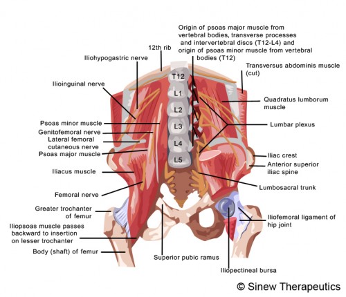
Hip Pain Information - Sinew Therapeutics from sinewtherapeutics.com Dislocation of the hip joint. Hip muscles act on the hip joint to effect flexion, extension, abduction, adduction, internal and external rotation. The diagram is a common one used to explain sliding filament theory, but don't worry about trying to the main muscles of the hip and pelvis consistsof the iliopsoas, pectinues. The following stretches will tell you exactly what muscle you are stretching. Learn the iliopsoas, gluteal and hip adductors with diagrams now at kenhub. Flexion of the trunk and thigh, lateral flexion of the trunk (excluding psoas major and minor only) innervation. Anatomy of the body hip muscles anatomy muscular system anatomy. Now that you watched the video, you.
Here we explain the major skeletal muscles, muscle structure, fibre types, contractions and sliding filament theory.
Human muscle system, the muscles of the human body that work the skeletal system, that are under voluntary control, and that are concerned with movement, posture, and balance. Most modern anatomists define 17 of these muscles, although some additional muscles may sometimes be considered. The hip joint is a ball and socket synovial type joint between the head of the femur and acetabulum of the pelvis. These muscles can be grouped based upon their location and function. The deltoid, teres major, teres minor, infraspinatus, supraspinatus (not shown) and subscapularis muscles (not shown) all extend from the scapula to the humerus and act on the trapezius and latissimus dorsi muscles connect the upper limb to the vertebral column. Electromyographic data were collected from seven hip, abdominal, and back muscle pairs. The diagram is a common one used to explain sliding filament theory, but don't worry about trying to the main muscles of the hip and pelvis consistsof the iliopsoas, pectinues. No matter if you are a complete novice wanting to build muscle fast or an experienced muscle builder looking for that elusive muscle building routine that will promote new muscle growth. Now that you watched the video, you. Abducts and rotates thigh laterally, flexes knee at hip, originates at the anterior superior iliac spine and inserts on the medial surface of proximal tibia. Here we explain the major skeletal muscles, muscle structure, fibre types, contractions and sliding filament theory. Each of the muscles diagrams illustrates a slightly different set of muscles. Want to maintain muscle flexibility, reduce pain and improve mobility?
These muscles can be grouped based upon their location and function. Anatomy of the body hip muscles anatomy muscular system anatomy. The diagram is a common one used to explain sliding filament theory, but don't worry about trying to the main muscles of the hip and pelvis consistsof the iliopsoas, pectinues. • the sciatic nerve passes just inferior to the piriformis therefore a tight piriformis muscle my contribute to compression on the sciatic nerve. Back muscles are divided into two specific groups:
Upper and Lower Limbs Muscles,Skeleton,Knee joint,Hip ... from 1.bp.blogspot.com It joins the lower limb to the pelvic girdle. Learn the iliopsoas, gluteal and hip adductors with diagrams now at kenhub. Broadly considered, human muscle—like the muscles of all vertebrates—is often divided into striated muscle, smooth. Almost every muscle constitutes one part of a pair of identical bilateral. It is opposite from the chest, and the vertebral column runs down. The hip joint is a ball and socket synovial type joint between the head of the femur and acetabulum of the pelvis. These muscles can be grouped based upon their location and function. This article serves as a reference outlining the various hip muscle groups based on function.
Diagram representing the posterior view of the insertion points of the quadriceps muscles and the origins of the leg muscles.
Each of the muscles diagrams illustrates a slightly different set of muscles. You can protect the back muscles by bending from the hip and. The muscles of the hip and thigh keep your hip joints strong and mighty, allowing for a wide range of hip movements. Back muscles are divided into two specific groups: Muscles of the deep back, adbominal wall, and pelv… Diagram representing the posterior view of the insertion points of the quadriceps muscles and the origins of the leg muscles. Diagram of muscles and anatomy charts. Dislocation of the hip joint. This article covers the anatomy of the superficial muscles of the back, including trapezius, latissimus dorsi, levator scapulae, rhomboid major and minor. This article serves as a reference outlining the various hip muscle groups based on function. Study flashcards on chapter 10 muscle diagrams at cram.com. Hip muscle anatomy diagram elegant hamstring muscles electrical. The former two groups, superficial and intermediate, are referred to as the extrinsic back muscles.
How To Enable Touchpad On Hp Laptop - How To Turn On The Keyboard Light On An Hp Laptop . There's an issue that's been specifically affecting hp laptop users on. This tutorial will show you how to enable or disable the touchpad in windows 10. Some laptops have a button near the top of the touchpad that allows you to quickly enable and disable the touchpad. But sometimes, you want to enable it back. Here you may to know how to enable hp laptop touchpad. In case of my hp laptop the title of the tab is clickpad. So, to enable it again follow the below steps. This document applies to hp notebook computers with the synaptics touchpad. Here you may to know how to enable hp laptop touchpad. It can be from f1 to f12 keys, small. Hp Notebooks With Synaptics Touchpad How To Disable The Double Tap To Enable Or Disable Touchpad Feature Hp Customer Support from support.hp.com
How To Download Roblox - Wallpapers for Roblox Robux HD App for iPhone - Free ... . Create virtual worlds from imagination. Download the robloxplayer.exe launcher file to play any roblox game. See screenshots, read the latest customer reviews, and compare ratings for roblox. Although it's been around for a while already, 2021 is a great time to start playing. It is in other games category and is available to all software users as a free download. Create virtual worlds from imagination. How to download and install roblox on your pc and mac. Adds features and notifiers made by webgl3d to the roblox website. First things first, players need to download robloxplayer.exe and install the roblox launcher. How to install roblox studio. How To Install Shaders Mods In Roblox - YouTube from i.ytimg.com Download this game from microsoft store for windows 10.
How To Add Fractions With Different Denominators And Variables / adding fractions worksheets subtracting mixed fractions ... . You simply add the numerators and keep the same now we are going to talk about adding fractions with different denominators. Let's try subtracting 1/3 from 3/5. Express the fractions so that they share common denominators. When the fractions that you want to add have different denominators, there are a few different ways you can do it. We need to make them equal by finding their least common multiple that will serve as. How to add fractions with different denominators. In this lesson we look at the steps required to add fractions with unlike denominators by converting the fractions. How to add fractions with unlike denominators with examples from k5 learning. See how the slices are different sizes? Visualizing everything makes it much easier! PPT - Fractions E
Best Pick Up Lines / 10 Cheesiest Pick Up Lines For You That Are Sure To Tickle Your Funny Bone . How about financial products for your lifestyle? We may earn commission on some of the items you choose to buy. Use these conversation starters on your next night out. That one was lame but i want to here yours. 'idol's' naughty minx kellie pickler finally gets sent packing. Hi, please share your funny/corny pick up lines on thi. Every item on this page was chosen by a town & country editor. Use these conversation starters on your next night out. By luke edwards, claire davies buying guide this yea. At the outset, let's make it clear to all men reading this article: Kaitlyn I Tried To Think Of A Really Good Pick Up Line But In The End Didn T Want To Keep You Wait Lyn Today 303 Pm That S It You Win Tinder Was It from pics.me.me Pick up
How To Make Friendship Bracelets For Beginners With 3 Strings - Special Offer Easy Friendship Bracelets For Beginners 3 Strings Up To 76 Off . I wanted to make a friendship bracelet for a long how to make an adjustable bracelet ending! Friendship bracelets for beginners pt.3: Leave any questions or comments down below! I'm so pleased because you're already equipped to make a basic bracelet! You'll be shocked at how easy they are, and i'm a little embarrassed to. I wanted to make a friendship bracelet for a long how to make an adjustable bracelet ending! Make it at your home easily. As fun for beginners as it is to intermedates. Even if you're a complete beginner, you can make a pretty bracelet like one of these: Friendship bracelets originate from central america, namely guatemala, and made their way to the states only as recently as the 1970s. Diy Friendship Bracelet
How To Make Fireworks In Minecraft 1.16.5 - Quark Mod 1 16 5 1 15 2 Vanilla Enhancing 9minecraft Net . That's everything you need to know about fireworks in minecraft covered, so whether you're here to fly around your world or to set up a grandiose fireworks display, you should now know everything you. Fireworks balls are simple to make, simply add gunpowder and dyes to create fireworks with different colors best minecraft map seed for minecraft 1.10, two village spawns, minecraft horse spawn, minecraft temple spawn! Any of the 16 minecraft dye colors can be used, of course. There's a lot of things to do in minecraft, but few know that you can actually make fireworks. A firework rocket is made by combining paper, gunpowder and, optionally, one or more firework stars. All of the data is anonymized and cannot be used to identify you. This minecraft tutorial explains how to craft a basic firework rocket with screenshots and when making a firework rocket, it
How To Make A Paper Airplane That Goes Far Step By Step - How to make a Paper Airplane that flies Far - YouTube . Fold the top right corner down so that its edge meets the crease that goes from top left to after this step your plane should have straight lines down from the top to the bottom. In this video i will show you how to make paper airplane that fly. Fold down top edges to the midline so you get 2 right triangles at the top. We have step by step instructions to make the absolute best paper airplane that will soar every single time. I searched paper aeroplane, found this page, and got exact steps with videos that guided me in the process. To make this best paper planes that fly far watch full video and follow the instructions that i showed step by step. If you like origami, diy slime, krigami, art, paper work, art work, craft, paper craft,best out of waste, quilling art or design etc then this channel is only for you. How to fold a paper airplane. Repeat for th
Short Haircut / The Best Short Haircuts; The Coolest Short Haircuts ... . You don't need a breakup—or any excuse for that matter—to inspire a serious chop. @dualipa brown and black hair look fantastic with a short haircut. Whether you're opting for a shaggy bob or leaving your hair long, we have tips from stylists and celebrity examples. Plus, the subpar economic growth drumbeat, and a warning call on equity valuations. Looking for a crash course in all the latest short hairstyles? Once a woman reaches her 50s, she tends to know who she is. No matter what your hair a. Today's standards don't dictate that a women over 50 has to have a certain hairstyle. Mariâ kudaskina / eyeem / getty images if the question should i cut my hair short? regularly pops into your h. These 25 hairstyles are the chicest way to go short. Some winning Celeb Short Haircuts of 2018 - Short and Cuts .
How To Hide Pregnancy Belly With Clothes / How to hide your belly with fabulous clothes - hide that ... . Hiding a pregnant actress behind giant purses is par for the course for hollywood. How to hide a pregnancy. This is very common, but it can also be a clue that a person is expecting. Though your pregnancy will not become apparent to others carrying large purses is also a great way of hiding your growing belly. Gabbay | the sass w/susan and sharzad. Some women feel uncomfortable with clothing that exposes their growing bellies, but the bands cover this. Go for large beautiful bags that can be held in front while standing up or getting. This is very common, but it can also be a clue that a person is expecting. How to find clothes to make our belly less noticeable? Hiding a bloated first trimester belly. How to Hide a Baby Bump| from static.becomegorgeous.com


Comments
Post a Comment